H37 was one of the first clinical isolates of the mycobacteria responsible for tuberculosis. The isolate formed rough-edged colonies and was highly virulent. The strain, H37Rv, has been used as a model for testing newly-discovered drugs against tuberculosis. For more than a century now, H37Rv has been continuously sub-cultured in labs.
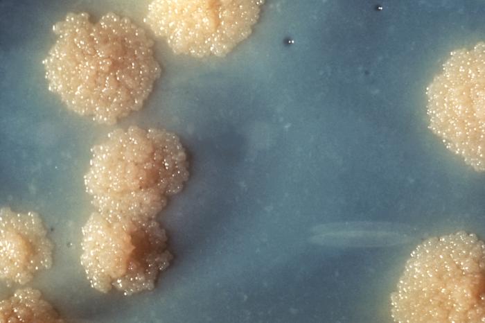
Colonies of Mycobacterium tuberculosis. Image: Dr. George Kubica via Wikimedia Commons
The favourable conditions in the lab made the strain progressively less virulent. However, strains now prevalent in patients mutated to become more virulent. And so H37Rv lost its prestige as a model for the mycobacterium that causes clinical tuberculosis.
How can we create a more appropriate virulent laboratory model of Mycobacterium tuberculosis?
Recently Vinay Kumar Nandicoori and team at NII, New Delhi collaborated with scientists from ICGEB, New Delhi to crack the problem. Instead of a culture medium, they needed a suitable host to grow the mycobacterium. The researchers chose guinea pigs because they are easy to breed and have a more robust immune system than that of mice.
The guinea pigs were infected with H37Rv. Two months later, the researchers harvested the lungs of the infected guinea pigs and homogenised the lungs. The homogenised lung lysate was plated on petri dishes to culture independent colonies of H37Rv.
Saba Naz, CCMB, isolated DNA from the bacilli. The fragmented DNA sequences were aligned to create a library with short overlapping sequences. Saba compared the whole genome sequence of Mycobacterium tuberculosis from the infected guinea pigs with that of the available standard H37Rv genome. Using bioinformatics tools such as the VarScan and SnpEff toolbox, she identified mutations at a single base pair level in the genome.
H37Rv in the guinea pigs acquired more mutation compared with the laboratory H37Rv strains. The result encouraged her to design an experiment. Saba used different strains, H37Rv and DNA repair deficient strain to challenge primary macrophages. To compare, she used H37Rv, as well as a combination of both strains. She infected the macrophages thrice to replicate real life scenarios, allowing time for mycobacteria to divide and mutate.
Late into the night, Saba counted the mycobacteria inside the macrophages. DNA repair deficient strains that had passed through the primary macrophages, she found, were greater in number. This indicated their evolutionary advantage of DNA repair deficient strain over H37Rv.
The scientists hope that the protocol will ensure a continuous supply of better cultures for laboratories working with tuberculosis bacteria. Strains with greater infectivity can now be used as models for testing drugs and to investigate the genetic basis of the virulence of the strain.
STAR Protocols 3 (4): 101804 (2022);
DOI: 10.1016/j.xpro.2022.101804
Reported by Bharati Swami
Product Development Cell-1, National Institute of Immunology
*This report was written in a workshop on science writing organised by NII

Congratulations Bharati
Aparna
LikeLike