Tear fluid is a valuable, non-invasive material for detecting the early signs of various diseases. So far, tears have been used to diagnose dry eye conditions. Now, researchers are discovering markers for various diseases in tears. However, methods for collecting, storing, and analysing tear samples vary widely, making it difficult to compare results or apply the findings in clinical settings.
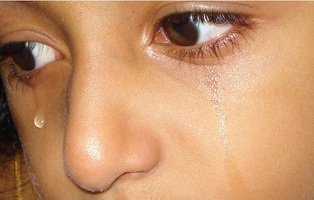
Image: Reji Jacob via Wikimedia Commons
To address these problems, the Tear Research Network, a network of clinicians, scientists, and researchers involved in tear fluid research and development, created an online survey. The survey aimed to map current practices related to research on tear fluid and to explore whether there is a shared need for standardised guidelines.
The survey was sent to researchers, eye doctors, and scientists worldwide. Participants were asked to reflect on their past five years of research practices, including how they collect, store, and analyse samples.
Researchers from fifty-nine labs across twenty-one countries completed the survey in one year. Most respondents worked in academic settings and were research scientists. Ophthalmologists were also in sizable numbers, since there are test kits available for detecting infections, inflammation and tissue degradation in the eyes, using tear fluid.
Around two-thirds of those who collected tear fluid had up to five years of experience in the field. Most researchers collected samples from both eyes. Only a small number used anaesthesia or any form of stimulation during collection.
Schirmer’s strips, a strip of Whatman paper that can be placed in the eye to assess the amount of tear, was the most common method used to collect tear fluid, followed by capillaries, special thin glass tubes. Though there were six other methods, only a few used them. Four-fifths of the people who used these methods did not use any normalisation strategy to account for variations in the volume of the fluid collected.
Three-fourths of the respondents collected more than six samples per month. One-fifth were collecting more than 20 samples each month. Samples were typically stored at minus eighty degrees Celsius within half an hour.
Although researchers froze the samples shortly after collection, they analysed the fluid only after several months. Among different analytes, proteins and peptides were the most commonly examined. Most researchers found the analysis of tear fluid the biggest challenge in their work.
There was a strong consensus on the need for clear standards in the field. Around ninety percent of those surveyed supported the creation of international guidelines, and eighty percent said they would be willing to change their current protocols if such standards were made available.
“The data clearly shows that the global community is ready for standardisation. With consistent practices, we can accelerate both research quality and its application in clinical care”, says Dr Swaminathan Sethu, Narayana Nethralaya Foundation, Bangalore.
A standardised protocol will significantly facilitate the identification of early diagnostic markers for various diseases, including multiple sclerosis, certain cancers, diabetic retinopathy, as well as Alzheimer’s and Parkinson’s disease.
But creating and testing shared protocols needs collaboration between researchers and clinicians globally. Creating awareness among stakeholders about the need for cooperation is the first step. This report is a part of that effort.
Contact Lens and Anterior Eye, 2025;
DOI: 10.1016/j.clae.2025.102388
Reported by K. Sri Manjari
Freelance science writer, Hyderabad
STEAMindiaReports: Free-to-use science news for Indian media
scienceandmediaworkshops
training to write better papers, reviews and project proposals

Leave a comment