Breast cancer is one of the most common cancers, affecting women worldwide. The quality of life and the survival of the patients depend largely on how early the disease is detected. In its early stages, breast cancer is often localized, and more responsive to treatment.
Traditional diagnostic tests, such as mammography, ultrasonography and core needle biopsy, take several days, generating stress in women suspected of having breast cancer. Moreover, these tests need equipment and skilled manpower that are not easily accessible.
How can we detect and diagnose breast cancer quickly and accurately to reduce the time and number of visits to clinics for getting test results and to initiate treatment at the earliest?
Recently, a team of researchers from the Netaji Subhash Chandra Bose Government Medical College and Hospital, Jabalpur came up with a way to quickly diagnose breast cancer, even in clinics with limited facilities.
Cells in cancerous tissues have nuclei with irregular shapes and rough surfaces whereas nuclei in normal tissues are more or less spherical and smooth. This can be detected using a good microscope with minimal training. But is this a dependable method to diagnose breast cancer?
From January to July, 2025, the researchers enrolled thirty female patients in their mid-thirties to late-sixties. The doctors of these women had detected lumps that suggested the possibility of breast cancer and had referred them to the medical college for tests to confirm.
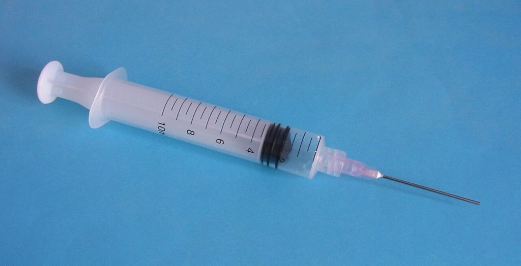
The research team in the medical college used ultrasound imaging and extracted biopsy samples from the lumps using syringes with 14G bore needles. Blood transfusions and ear piercing are often done using needles with higher bore dimensions.
Image: Biggishben via wikimedia commons
From each patient, six to eight small samples were taken. Immediately after the biopsy, the tissue samples were gently touched onto clean glass slides and rolled to imprint the cells and make smears for microscopic examination.
“Preparing the slides using touch imprint did not compromise the morphology of the core samples”, says Dr Manasi Nema, Netaji Subhash Chandra Bose Government Medical College and Hospital, Jabalpur
The doctors compared the results from histologically detected cases with the results from other tests. One woman had no breast cancer. In twenty-eight cases, the results from the other tests agreed with the results from the touch imprint cytology test. Only in one case, the result from touch imprint cytology turned out to be a false positive..
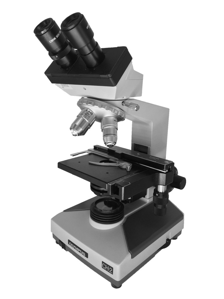
Images: Biggishben and Sarah Greenwood via Wikimedia Commons
The results from the comparison suggest that the touch imprint cytology test can reduce the number of cases referred to major hospitals from smaller clinics. Even in resource limited rural areas, doctors can easily use touch imprint cytology, since it requires only a microscope and minimal experience and skills to distinguish normal from cancerous cells.
Women suspected of having breast cancer due to the detection of lumps in their breasts can get the results in a few hours. The anxiety of many days of waiting for test results can be eliminated.
Only those whose tests are positive need to be sent for confirmatory tests before initiating treatment.
“Touch imprint cytology using core needle biopsy is a valuable tool, but underused in health care settings in India”, remarks Dr Sanjay Kumar Yadav, Netaji Subhash Chandra Bose Government Medical College and Hospital, Jabalpur.
DOI: 10.1177/00494755251383963;
Tropical Doctor, 56 (1): 83–85 (2026)
Reported by Sanghamitra Deobhanj
Freelance science writer, Cuttack
The reports on this site are free-to-use for Indian media houses.
This report was written in a workshop for science communicators organised by scienceandmediaworkshops.
Scienceandmediaworkshops offers similar capacity building exercises
for Indian researchers, scientists and science faculty
to write scientific manuscripts, project proposals and project reports.
The next workshop for Indian research community starts on 4th April 2026

Leave a comment